
NL Journal of Dentistry and Oral Sciences
(ISSN: 3049-1053)
Functional and Aesthetic Rehabilitation of Distal Extension Edentulous Arches with Extra-Coronal Attachments: A Clinical Case Utilizing the PreciVertix System
Author(s) : Arpit Sikri, Vaishali Kalra, Jyotsana Sikri. DOI : 10.71168/NDO.01.01.112
Abstract
Rehabilitating Kennedy Class I and II cases, particularly when opposing a partially edentulous arch, presents notable challenges due to retention difficulties. While implant-supported prostheses are often the ideal solution, systemic health limitations can sometimes preclude their use. Conventional clasp-retained partial dentures provide an alternative, but they lack aesthetic appeal. In contrast, extra-coronal attachmentretained prostheses offer a more visually pleasing and accessible option. Despite the long-standing availability of dental attachments, their advantages remain underutilized in clinical practice. Attachments can significantly enhance comfort, aesthetics, functionality, and patient satisfaction, especially in cases of long-span edentulism where implants or fixed partial dentures are unsuitable. For patients with unilateral or bilateral posterior tooth loss, attachment-retained cast partial dentures provide a viable solution. Precision attachments are designed to balance stability and aesthetics while minimizing excessive forces on the abutment teeth and ridges during use. Semi-precision attachment systems are widely valued for their ability to provide stability, retention, and aesthetics in removable partial dentures, thereby enriching traditional treatment options. The present case demonstrates the use of an extra-coronal precision attachment (Preci-Vertix attachment system) to restore maxillary bilateral distal extension edentulous spans. Keywords: Attachment Retained Denture, Bilateral Distally Extended Removable Partial Denture, Bilateral Posterior Region Teeth, Cast Partial Dentures, Distal Extension Attachments, Distal Extension Cases, Distal Extension Removable Partial Denture, Preci-Vertix, Precision Attachments, Semi-Precision Attachments, Snap Fasteners.
Introduction
Since the early 20th century, precision and semi-precision attachments have served as connectors that offer stability and retention in prosthodontic applications [1]. These attachments consist of two parts: one component attaches to a tooth, root, or implant, while the other connects to the prosthesis [2].
Attachments can be categorized in two main types. Precision attachments are prefabricated from durable alloys and are rigidly welded to the prosthesis, ensuring a strong and reliable connection. Semi-precision attachments, however, are cast from plastic patterns, making them slightly less precise but more resilient and cost-effective, suiting particular clinical needs [3].
Attachments are also classified by their location. Intracoronal attachments are positioned within the crown’s contour, requiring substantial tooth preparation, while extracoronal attachments are placed outside the crown, reducing the amount of tooth reduction necessary. Common semi-precision extracoronal attachment systems include Preci-Vertix and ERA attachments [4]. Bar attachments, such as Preci Bars or Hader Bars, span multiple abutments across edentulous spaces, providing stability. Stud attachments, which include radicular and intraradicular types, directly attach to roots or root contours for alternative support [5].
Attachments can be either rigid or resilient. Rigid attachments are commonly used in fixed prostheses to limit movement, while resilient attachments allow slight movement, making them ideal for tooth-tissue-supported dentures by helping to distribute forces and reduce stress on the abutments [6].
Various mechanisms achieve retention in attachments, including frictional retention, mechanical interlocking, or magnetic attraction. Choosing the right attachment system depends on factors like the type of prosthesis, intended function and location, retention needs, space constraints, and cost [7]. In India, the wide availability of attachments, their affordability, and the expertise of skilled technicians have boosted their popularity in treating partially edentulous patients.
Attachments play a critical role in rehabilitating distal extension cases, where missing posterior teeth create a freeend saddle [8]. These cases, particularly when opposing a partially edentulous arch, present unique challenges. Fixed prostheses may not be feasible due to long cantilever spans, and while implant solutions are effective, they can be expensive. Conventional cast partial dentures can compromise aesthetics due to visible metal clasps. In these cases, precision and semi-precision extra coronal attachments provide a superior option, offering enhanced aesthetics and functionality without the visible drawbacks of traditional clasps [9].
Research shows a five-year survival rate of 83.35% and a 20-year survival rate of 50% for attachment-retained dentures [10]. In prosthodontics, precision attachments used in removable partial dentures and other prosthetic designs offer significant functional and aesthetic advantages, adding a new dimension to treatment options. This paper discusses a case where a maxillary Kennedy Class I condition was restored with semi-precision attachments, opposing a partially edentulous mandibular arch.
Case Report
A 45-year-old male patient presented to the Department of Prosthodontics, Crown & Bridge, and Oral Implantology at Bhojia Dental College & Hospital, Baddi, Solan, Himachal Pradesh, India, with a chief complaint of needing capping for root canal-treated teeth in the upper front region, along with replacement of partially missing teeth in both the upper and lower anterior and posterior regions. Clinical examination revealed remaining natural teeth from 13 to 23. An OPG confirmed generalized bone loss in both the maxillary and mandibular arches.
In terms of his medical history, there were no relevant concerns reported. The patient described his attitude towards treatment as philosophical and adhered to a vegetarian diet. He brushes his teeth once a day and denied any history of smoking, alcoholism, tobacco chewing, bruxism, or clenching. The patient’s primary expectation from treatment was to achieve proper function.
General examination revealed an average-built individual with a well-coordinated gait who was oriented to person, place, and time. His vital signs were normal. Upon extraoral examination, the form of his face was square tapering, with a straight profile. His facial symmetry was noted as symmetrically asymmetrical, and the facial height appeared normal. The temporomandibular joint (TMJ) examination showed no tenderness or clicking sounds, and there were no palpable lymph nodes.
Intraorally [Figure 1], the patient had undergone multiple extractions due to caries, including the removal of teeth 12, 14, 15, 16, 17, 22, 24, 25, 26, 27, 36, 37, 45, 46, and 47 over the past six months. Endodontic treatment had been performed on teeth 11, 13, 21, 23, and 35. Tooth 23 was palatally positioned, resulting in a cross-bite, while cervical abrasions were observed on teeth 34, 35, 41, 43, and 44.
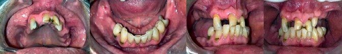 Figure 1 – Intraoral examination
The patient’s general health status was assessed as fair, with no abnormalities detected in the lips, cheeks, tongue, floor of the mouth, palate, or temporomandibular joint (TMJ), although mild gingivitis was noted. The occlusal pattern was characterized by canine guidance.
The diagnosis for this patient was determined to be a partially edentulous case, classified as Kennedy’s class I in both the maxillary and mandibular arches.
For treatment planning, several options were considered. These included implant-supported fixed prostheses for the upper and lower missing teeth, cast partial dentures, and precision attachment retained fixed-removable prostheses for both arches. Additionally, porcelain fused to metal (PFM) crowns with precision attachment retained fixed-removable prostheses were proposed for the upper arch, along with a flipper for the lower arch. Other options included PFM crowns followed by cast partial dentures for both the upper and lower arches, and all-ceramic crowns for the upper front teeth, also followed by cast partial dentures for both arches. Extractions of all upper arch teeth were also discussed, followed by implant retained prostheses or complete dentures.
For treatment options, implant-supported prostheses were not considered viable due to financial constraints. Instead, a less invasive treatment plan was devised, focusing on a tooth-supported fixed prosthesis extending from the upper right canine to the left canine, combined with semi-precision attachments for an attachmentretained removable dental prosthesis (excluding a cast partial denture).
PFM crowns with precision attachment retained fixed-removable prostheses were selected for the upper arch, along with a flipper for the lower arch. The choice of retainers included PFM full coverage crowns for teeth 13 to 23. The materials of choice comprised bis-acryl composite (Coltene Whaledent Cooltemp) for temporary restoration on teeth 13 to 23, with porcelain fused to metal crowns for final restoration on the same teeth. Luting cements were specified, with zinc phosphate cement for the cementation of provisionals and glass ionomer cement (GIC) for the permanent cementation of the crowns. Patient education focused on oral hygiene instructions, including the use of dental floss, brushing around the attachments, and keeping the prosthesis soaked in water to prevent cracking or distortion.
Procedure
1. Diagnostic Stage: The patient’s diagnostic casts [Figure 2] were fabricated, and the treatment planning process was completed. Denture bases were constructed using self-cure acrylic resin on the casts, and occlusal rims were formed with modeling wax. The orientation jaw relation was recorded with the aid of a facebow [Figure 3]. The vertical dimension and tentative centric relation were established and transferred to a semi-adjustable articulator [Figure 4]. Subsequently, a mock-up wax pattern was designed on the diagnostic cast [Figure 5].
 Figure 2 – Diagnostic impressions and casts
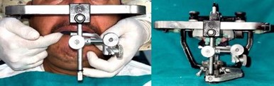 Figure 3 – Facebow transfer
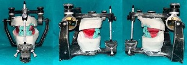 Figure 4 – Centric relation record and diagnostic mounting
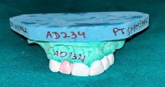 Figure 5 – Mock-up pattern
2. Tooth Preparation and Impressions: Tooth preparation was performed on the other diagnostic cast, followed by the creation of a check impression and check cast [Figure 6]. Atraumatic tooth preparation [Figure 7] was carried out with supragingival margins for teeth 13–23. A putty index was made from the mock wax-up cast, and provisional restorations were then fabricated [Figure 8]. Impressions of the maxillary and mandibular arches were taken using the putty-wash technique [Figure 9]. The final maxillary and mandibular casts are shown in Figure 10. Tentative jaw relations were recorded [Figure 11]. A facebow record was taken on the prepared maxillary cast and transferred to the semi-adjustable articulator [Figure 12].
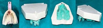 Figure 6 – Check impression and cast
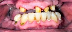 Figure 7 – Tooth preparation
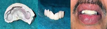 Figure 8 – Provisionalization
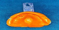 Figure 9 – Final impression
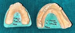 Figure 10 – Final cast
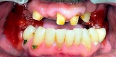 Figure 11 – Jaw relation record
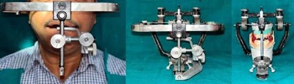 Figure 12 – Facebow record and mounting on a semi-adjustable articulator
3. Framework and Attachment: The prefabricated castable attachments (Preci-Vertix) were incorporated into the wax pattern for the metal copings of the fixed partial denture (FPD). These extra-coronal attachments, with elastic retention, functioned as stress breakers and absorbed excessive forces.
4. Casting and Fitting: The metal copings were cast using the lost-wax technique and checked intraorally for fit [Figures 13 & 14]. A pick-up impression was made using polyvinyl siloxane material to ensure precise fitting of the attachments. The facebow record was taken and transferred, and the centric jaw relation record was completed after confirming the metal try-in in the patient’s mouth [Figure 15].
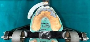 Figure 13 – Metal trial with male attachment (extraoral)
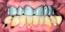 Figure 14 – Metal trial with male attachment (intraoral)
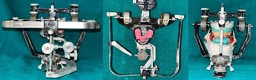 Figure 15 – Facebow transfer and mounting on a semi-adjustable articulator
5. Definitive Prosthesis: After applying the ceramic and completing all associated procedures on the master cast, the Preci-Vertix Ceka plastic housing (female attachment) was placed over the male attachment of the metal [Figure 16]. The teeth arrangement for both the maxillary attachment-retained partial denture (without CPD framework) and the mandibular RPD was completed, followed by a wax trial to evaluate occlusion, phonetics, and aesthetics. The dentures were then processed, and the female attachments were incorporated into the maxillary denture base, allowing for future replacement if necessary.
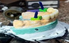 Figure 16 – Attachment of Preci-Vertix Ceka plastic housing
6. Final Insertion: A bisque trial was conducted in the patient’s mouth [Figure 17]. During the cementation of the fixed partial denture (FPD), the attachment-retained partial denture (without CPD framework) was aligned extra-orally, ensuring proper insertion of both the fixed and removable prostheses [Figure 18]. A flexible partial dental prosthesis was provided for the mandibular arch [Figure 19]. The prostheses were then inserted [Figure 20], offering the patient both an aesthetic and functional solution [Figure 21].
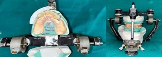 Figure 17 – Bisque trial
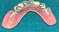 Figure 18 – Maxillary removable prosthesis with incorporated female component
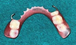 Figure 19 – Mandibular flexible denture
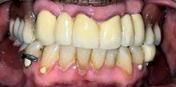 Figure 20 – Final prostheses
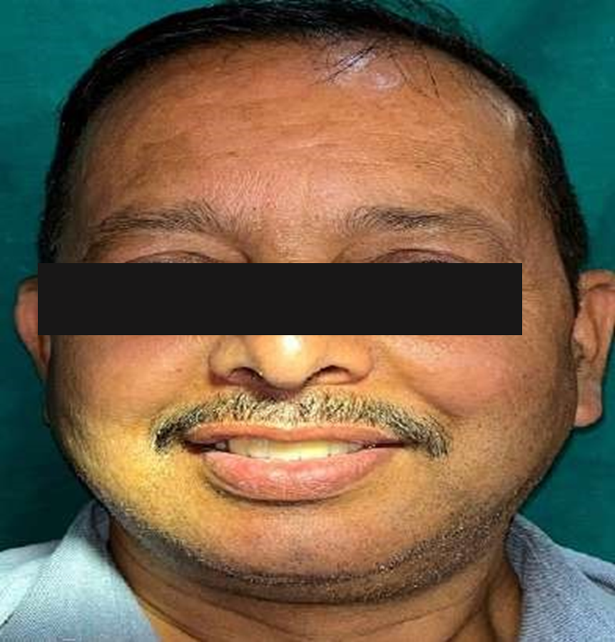 Figure 21 – Happy patient (Post-op view)
This case demonstrates the use of semi-precision attachments in prosthodontics and selecting non-invasive prosthetic options tailored to the patient’s overall health.
Discussion
Rehabilitating patients with distal extension partially edentulous arches, such as those in Kennedy Class I and II cases, poses significant clinical challenges. A primary difficulty stems from the absence of a posterior abutment tooth, which compromises the retention, support, and stability of removable partial dentures (RPDs) [11]. Conventional cast partial dentures (CPDs) are often used but can be aesthetically and functionally limited due to visible clasps, particularly in anterior aesthetic zones [12]. Furthermore, the saddle areas of CPDs are prone to rotational movements because of insufficient support from the distal edentulous ridge.
While implant-supported fixed partial dentures (FPDs) are generally considered the ideal solution for these cases, they may be contraindicated due to the patient’s systemic health, financial constraints, or inadequate bone volume [13]. In situations where implants are not viable, precision and semi-precision attachments present a promising alternative. These attachments, commonly used with CPDs, enhance aesthetics by eliminating visible clasps and improve functional outcomes by serving as stress breakers [14].
Precision attachments comprise two components: one attaches to the prosthesis, while the other connects to an abutment tooth, root, or implant. This configuration allows for a connection that integrates features of both fixed and removable prostheses [15]. In distal extension cases, the extracoronal type of attachment is preferred as it accommodates vertical and lateral movements, distributing occlusal forces away from the abutments and minimizing the risk of damage to the supporting structures. It is advisable to splint the distal abutment with the mesial tooth for additional support and to incorporate lingual shoulders or bracing arms to enhance stability [16].
Preci-Vertix is a bolt-type semi-precision extracoronal attachment designed for removable partial dentures (RPDs), offering several advantages, including simple fabrication procedures and servicing, improved esthetics, cost-effectiveness, and excellent patient comfort [17]. The matrixes of the precision attachment system are made from various polymers and are available in three color-coded retention values, which correspond to their frictional features: yellow for standard retention, white/green for decreased retention, and red for increased retention [18].
These attachments operate based on the Broken Stress philosophy, allowing for vertical movement while reducing stress transfer to abutments. Staubli has classified attachments into six categories: Class 1a, which includes solid, rigid, non-resilient attachments; Class 1b, comprising solid, rigid, lockable attachments with a U-pin; Class 2, which features vertical resilient attachments; Class 3, including hinge resilient attachments; Class 4, which consists of both vertical and hinge resilient attachments; Class 5, encompassing rotational and vertical resilient attachments; and Class 6, which includes universal, omniplanar attachments [19,20].
Precision attachment partial dentures do not rely on clasps for retention, thereby eliminating the wedging effect of clasps and facilitating a more favorable distribution of horizontal forces [21]. These attachments effectively tie abutment teeth together, limiting excessive movement. In the retainer design, centrals, laterals, and canines were included as abutments to provide the extra leverage required, whereas conventionally only one or two abutments would typically suffice [22].
The selection of abutments plays a critical role in the attachment-retained prosthesis [23]. Canines are often chosen as anchors due to their proprioceptive nature, favorable form and strategic position, as well as the wider periodontal fixation area they provide. Partial dentures constructed using canines as abutments are less prone to breakage and are effective in distributing temperature changes [24].
Preci-Vertix attachments are a commonly utilized option, offering prefabricated components that can be cast directly with the wax pattern, thereby simplifying the fabrication process and improving accuracy [25]. However, the small and technique-sensitive components of the attachment system necessitate precise laboratory work. Over time, wear and tear may require the periodic replacement of certain parts, such as the nylon caps in the female component [26].
The advantages of precision attachments include improved retention, enhanced aesthetics (due to the absence of visible clasps), and increased support and stability for distal extension RPDs [27]. However, their success is heavily reliant on the clinician’s skill, the accuracy of the laboratory work, and the patient’s adherence to followup care. Despite potential challenges in fabrication and maintenance, precision attachments remain a highly effective and aesthetic option for rehabilitating distal extension edentulous arches when implant therapy is not feasible [28].
However, there were several issues encountered with the case. Condensation silicone was used for making the maxillary diagnostic impression, and Broadrick’s flag was not utilized to establish the occlusal plane. There was also a discrepancy in shade matching with respect to the 35 PFM crown. Additionally, due to economic reasons, attachments were incorporated into the flipper instead of utilizing cast partial dentures. Finally, the upper and lower midlines were not aligned in the final prosthesis [29,30,31].
Conclusion
The incorporation of precision and semi-precision attachments in removable partial dentures (RPDs) can significantly enhance patient comfort, aesthetics, and functional outcomes, particularly in long-span edentulous cases where fixed prostheses (FPDs) or implants are not feasible. Although the fabrication process may be complex and time-consuming, a strong understanding of prosthodontic principles and sound clinical judgment enable clinicians to effectively integrate these attachments into routine practice.
There are several advantages to using attachments. First, they improve aesthetics by eliminating unsightly clasps, which enhances patient acceptance and satisfaction. Second, they provide superior retention, with systems like the Preci-Vertix offering excellent fit and the option to replace retentive caps over time to maintain or improve retention. Third, they present a cost-effective alternative to surgical solutions, such as implants, making them an affordable treatment option for distal extension cases. Additionally, hybrid prostheses that combine fixed and removable features provide stability, improved occlusal load distribution, and aesthetic appeal.
However, challenges exist. The fabrication of precision attachments is highly technique-sensitive, requiring meticulous attention to detail during both the clinical and laboratory phases. Furthermore, regular follow-ups, typically every three months, along with preventive maintenance, are crucial for long-term success, as components may need periodic adjustment or replacement.
In distal extension cases, the clinical application of attachments effectively bridges the gap between fixed and removable prosthetics. These attachments provide both stability and the ability to enhance retention without surgical intervention, making them particularly suitable for patients with Kennedy Class I and II edentulous arches.
In conclusion, precision attachment-retained partial dentures strike a balance between traditional and modern prosthodontic techniques. With careful treatment planning and attention to patient-specific needs, they can provide a functional, aesthetic, and comfortable solution for complex edentulous cases. The success of this approach relies on continuous evaluation, periodic maintenance, and adherence to preventive strategies to ensure long-lasting results.
References
1. McCracken, W. S. (1999). McCracken’s removable partial denture prosthodontics (12th ed.). Mosby.
2. Bakers, J. L., & Goodkind, R. J. (1981). Precision attachment removable partial dentures. Mosby.
3. Karr, A. B., & Brown, D. T. (Year). McCracken’s removable partial denture prosthodontics. Mosby Elsevier.
4. Burns, D. R., & Ward, J. E. (1990). Review of attachments for removable partial denture design: I. Classification and selection. The International Journal of Prosthodontics, 3(1), 98-102.
5. Burns, D. R., & Ward, J. E. (1990). A review of attachments for removable partial denture design: II. Treatment planning and attachment selection. The International Journal of Prosthodontics, 3(2), 169-174.
6. Angadi, P. B., Aras, M., William, C., & Nagaral, S. (2012). Precision attachments: Applications and limitations. Journal of Evolution of Medical and Dental Sciences, 1(6), 1113-1121.
7. Gupta, N., Bhasin, A., Gupta, P., & Malhotra, P. (2013). Combined prosthesis with extracoronal castable precision attachments. Case Reports in Dentistry.
8. Jain, A. R. (2013). A prosthetic alternative treatment for severe anterior ridge defect using fixed removable partial denture Andrew’s bar system. World Journal of Dentistry, 4(4), 282-285.
9. Jain, R., & Aggarwal, S. (2017). Precision attachments: An overview. Annals of Prosthodontics and Restorative Dentistry, 3(1), 6-9.
10. Munot, V. K., Nayakar, R. P., & Patil, R. (2017). Prosthetic rehabilitation of mandibular defects with fixed RPD prosthesis using precision attachment: A twin case report. Contemporary Clinical Dentistry, 8(3), 473-478.
11. Vermeulen, A. H., Keltjens, H. M., Van’t Hof, M. A., & Kayser, A. F. (1996). Ten-year evaluation of removable partial dentures: Survival rates based on retreatment, not wearing, and replacement. Journal of Prosthetic Dentistry, 76(3), 267-272.
12. Viennot, S., Dalard, F., Malquarti, G., & Grosgogeat, B. (2006). Combination fixed and removable prostheses using a CoCr alloy: A clinical report. Journal of Prosthetic Dentistry, 96(2), 100-103.
13. Zinner, I. D., Miller, R. D., Parker, H. M., & Panno, F. V. (1989). Prefabricated metal intracoronal semi-precision attachments for removable partial dentures. International Journal of Prosthodontics, 2(4), 357-364.
14. Weaver, S. M. (1938). Precision attachments and their advantages in respect to underlying tissues. Journal of the American Dental Association, 25(8), 1250-1259.
15. Wagner, B., & Kern, M. (2000). Clinical evaluation of removable partial dentures ten years after insertion: Success rates, hygienic problems, and technical failures. Clinical Oral Investigations, 4(2), 74-80.
16. Nigam, A., Singh, A., Shekhar, A., & Gupta, H. (2013). Precision attachments: An overview. Journal of Dental and Facial Sciences, 2(4), 41-44.
17. Bambara, G. E. (Year). Precision and semi-precision attachments. In Freedman, G. A. (Ed.), Contemporary esthetic dentistry (1st ed., Chapter 25, p. 575). Elsevier.
18. Makkar, S., Chhabra, A., & Khare, A. (2011). Attachment retained removable partial denture: A case report. International Journal of Clinical Dentistry Sciences, 2(2), 39-43.
19. Konwar, A. K., Parameswari, D., & Annapoorni, H. (2020). Removable denture options in rehabilitation of missing dentition: A series of case reports. Annals of Prosthodontics and Restorative Dentistry, 6(2), 110-113.
20. Kumar, R. D., Parameswari, B. D., & Annapoorni, H. (2020). Rehabilitation of partially edentulous patients using precision attachment denture: A case report. IP Annals of Prosthodontics and Restorative Dentistry, 6(3), 162-166.
21. Mukhopadhyay, N., Das, S. L., Sen, U. K., Mondal, S., & Banerjee, S. (2022). Functional rehabilitation of mandibular distal extension partial edentulous arch combined with maxillary complete edentulism. International Journal of Clinical Medicine and Case Reports, 19(3), 002.
22. Patil, A. P., Pawar, A. V., & Patil, R. (2020). Unique, cost-effective and retentive removable prosthesis to rehabilitate long span Kennedy’s Class I edentulism with custom attachment system: A case report. Journal of Contemporary Dental Practice, 21(2), 215-218.
23. Turagam, N., Mudrakola, D. P., Yelamanchi, R. S., Deepthi, M., & Natarajan, M. (2019). Esthetic clasp cast partial denture. Journal of the International Society of Preventive & Community Dentistry, 9(1), 94-98.
24. Patil, R., & Shetty, O. (2019). Prosthetic rehabilitation using extracoronal attachments. International Journal of Dental Research, 4(1), 5-8.
25. Arita, S., Gonda, T., Togawa, H., Maeda, Y., & Ikebe, K. (2020). Influence of mandibular free-end partial edentulism on the force exerted on maxillary anterior teeth. Journal of Prosthodontic Research, 64(4), 454-459.
26. Shakeel, S. K. (2013). Removable prosthesis using extracoronal precision attachment. Gulf Medical Journal, 2, 126-129.
27. Glossary of prosthodontic terms (9th ed.). (2016). Journal of Prosthetic Dentistry, 117, 13-14.
28. Ceka Preci line. (n.d.). About Us. Retrieved from www.ceka-preciline.com.
29. Sterngold Attachments. (n.d.). Retrieved from www.sterngold.com.
30. Becerra, G., et al. (1987). A classification of precision attachments. Journal of Prosthetic Dentistry, 56, 322-327.
31. Prashad, K. D., et al. (2016). A simplified approach to semi-precision attachment. NUJHS, 6, 51-57.
This article licensed under the Creative Commons Attribution 4.0 International License CC-BY 4.0., which permits unrestricted use, distribution, and reproduction in any medium, provided the original author and source are properly credited.