
NL Journal of Medical and Pharmaceutical Sciences
(ISSN: 3108-0502)
Neurogenesis in the Hippocampus and its Influence on Cognitive and Emotional Abilities
Author(s) : Elizaveta I Bon*, Maksimovich N Ye, Zimatkin S M, Yusko E V. DOI : 10.71168/NMP.01.01.103
Abstract
The neurogenesis of the hippocampus in adults plays a key role in cognitive, emotional, and behavioral functions, sparking discussions about its evolutionary significance. Differences between neurogenesis during development and in adulthood are emphasized, suggesting that they serve different functions: the constitution and lifelong adaptation of cognitive and emotional behavior. This distinction also indicates varying vulnerabilities to mental disorders depending on the life stage. We propose new research directions focused on the impact of hippocampal neurogenesis on mental disorders, which could deepen the understanding of risk factors and adaptation mechanisms. Keywords: hippocampus, neurons, neurogenesis.
Introduction
In most mammals, new neurons are formed not only during the embryonic period but also after birth. Soon after the discovery of neurogenesis in adults, it was noted that the addition of new neurons affects cognitive functions, particularly memory. Similarly, late neurogenesis also impacts affective functions, although this fact has been acknowledged with difficulty. Current data show that new neurons can arise not only from stem cells but also from neuroblasts, which exhibit prolonged maturation and may participate in functional brain circuits under certain signals. The importance of adding new neurons to brain networks in adults is discussed, especially in the context of affective outcomes [1]. It is also highlighted that adult neurogenesis could be a limited cellular process that integrates factors from both internal and external environments to modulate brain functions. Research on neurogenesis in humans offers new insights into people as mammals. This ability could serve as a foundation for treating mental disorders, cognitive impairments, and age-related diseases. The concept of an adult brain that consistently incorporates new neurons marks a significant advancement in creating innovative therapeutic strategies for addressing mental health disorders linked to high rates of morbidity, mortality, and societal expenses [2].
Objective
To investigate the influence of new neurons in the hippocampal formation on the affective functions of the brain in adult mammals, including humans, and to assess the potential of neurogenesis as a means to improve mental health and treat cognitive disorders
Methodology
The foundation of this study was a review of contemporary foreign and domestic literature on this topic.
Results and Discussion
The hippocampus is made up of closely packed cells organized in a ribbon-like formation that runs along the inner walls of the lower horns of the brain’s lateral ventricles, extending from front to back. The two halves of the hippocampus are linked by commissural nerve fibers.
The hippocampal formation consists of the “proper hippocampus,” which includes fields CA1, CA2, and CA3, along with the dentate gyrus and the subiculum [2]. The proper hippocampus is further divided into proximal large-cell and distal small-cell areas, with fields CA3 and CA2 corresponding to the large-cell area, and CA1 to the small-cell area. The ventricular surface of the hippocampus is referred to as the alveus. The alveus is a delicate layer of white matter made up of the axons from the pyramidal cells in the hippocampus, and it is topped with a layer of ependymal cells. The fibers of the alveus come together at the inner border of the hippocampus, creating the fimbria of the hippocampus, which subsequently develops into the fornix [3]. According to modern histological nomenclature, the proper hippocampus is distinguished into three layers:
- Molecular (molecular layer), which consists of the eumolecular (eumolecular substrate), lacunar (lacunar substrate), and radial (radial substrate) sublayers.
- Pyramidal layer (stratum pyramidale).
- Oriens layer (stratum oriens) (Figure 1).
The structure of these layers is generally consistent across all fields of the hippocampus (Table 1) [5,6,7].
Table 1: Neuronal and transmitter organization of the hippocampus.
| Name of the Neuron | Layers (Sub-Layers) of the Cortex | Afferent Innervation | Efferent Innervation | Mediator |
|
Non-pyramidal interneurons. |
Molecular (stratum molecular). |
Axons of neurons from the entorhinal cortex and thalamic nuclei, non-pyramidal interneurons in the molecular and marginal layers. | Non-pyramidal interneurons in the molecular and marginal layers, dendrites of pyramidal neurons. |
GABA |
| Non-pyramidal interneurons. | Lacunar sublayer (substratum lacunosum). | Non-pyramidal interneurons in the molecular and marginal layers. | –//– | GABA |
| Non-pyramidal interneurons. | Radial sublayer (substratum radiatum). | Non-pyramidal interneurons of the molecular and marginal layers. | –//– | GABA |
|
Pyramidal neurons. |
Pyramidal (stratum pyramidale). |
Axons of granule neurons of the dentate gyrus, Schaffer collaterals, axons of basket cells, and all other neurons of the molecular and marginal layers. |
Schaffer collaterals, neurons of the entorhinal cortex, and basket neurons. |
Acetylcholine. |
| Basket neurons. | Pyramidal. | Axons of pyramidal neurons. | Pyramidal neurons. | GABA |
| Trilaminar neurons. | Pyramidal. | –//– | Neurons of the subiculum | GABA |
| Candelabrum cells. | Pyramidal. | –//– | Pyramidal neurons. | GABA |
| Non-pyramidal interneurons. | Stratum oriens. | Non-pyramidal interneurons of the molecular and stratum oriens layers. | Pyramidal neurons. | GABA |
In the molecular layer, there are three types of non-pyramidal GABAergic neurons [7]. In the eumolecular sublayer, there exists a cluster of fibers that extend from the subiculum, concluding afferent pathways originating from the entorhinal cortex and mid-thalamic nuclei. Meanwhile, in the lacunar sublayer, axons travel from the hippocampus to the subiculum.
In the CA3 region, unlike in the CA2 and CA1 regions, there is a narrow acellular zone situated just above the layer of pyramidal neurons, where the axons of granule cells (known as the stratum lucidum) are located. At the far end, these fibers create a curve that marks the boundary between the CA3 and CA2 fields.
The substratum radiatum includes nerve fibers that establish connections between the neurons of the CA3 and CA1 fields. The pyramidal layer is the primary layer of the hippocampus. It contains pyramidal, basket, tri-laminar neurons, and chandelier cells. The dendrites of pyramidal cells are directed towards both the molecular and the marginal layers [8]. Lorente de No (1934) observed variations in the dendritic structure of pyramidal cells across distinct regions of CA3 and CA1. This led him to further categorize these areas into three subregions: CA3 a, b, c and CA1 a, b, c. [10].
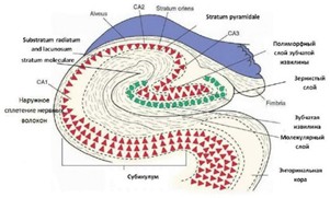 Figure 1: Diagram of the neuronal organization of the rat hippocampus (according to data from the US National Library of Medicine) [14].
The thin, mostly acellular marginal layer consists of the basal dendritic branches of pyramidal neurons, along with the cell bodies and dendritic branches of polymorphic (non-pyramidal) interneurons. In the hippocampus itself, eight types of neurons are distinguished. The main ones, pyramidal neurons, are cholinergic, while the others are GABAergic. (Table 1) Pyramidal neurons can be found in the pyramidal layer [11,12]. Their dimen- sions and arrangement in CA3 vary sequentially based on their location along the CA3 axis. Cells in the proximal region, situated close to the dentate gyrus, exhibit the shortest dendritic branches. In contrast, neurons located in the distal area of the CA3 field (adjacent to CA2) possess the longest dendritic branches. CA2 features a diverse group of neurons, including those with extensive dendritic branching comparable in size to neurons found in the distal region of CA3. Additionally, it includes cells with smaller dendritic branches that are similar to pyramidal neurons in CA1. Between 42% and 51% of the basal dendrites of CA3 pyramidal neurons are situated in the stratum oriens. Additionally, 34% of the dendritic branches from CA1 neurons also reach into this layer. Moreover, 18% of the apical dendrites from the same areas extend into the stratum moleculare and lacunosum. Alongside pyramidal neurons, the pyramidal layer of the hippocampus contains a diverse group of basket cells that vary in size and shape. They possess both apical and basal dendritic branches [13,14]. The axons of basket neurons spread horizontally from the cell bodies, creating basket-like structures that synapse with the cell bodies of pyramidal neurons in the hippocampus. Basket neurons receive excitatory signals from pyramidal neurons and have an inhibitory influence on them. Pyramidal cells produce recurrent excitation, a key process involved in the formation of memories. (Table 1) There are various kinds of non-pyramidal interneurons. They can be found in the molecular and outer layers. The vast majority of them are considered local circuit neurons. The dendrites of hippocampal interneurons reach into the stratum oriens, whereas their axons establish synapses in the stratum moleculare. These cells form synapses with the dendrites of pyramidal neurons, leading to inhibitory effects on them. (Table 1) Certain interneurons in the CA1 region exhibit significant axonal branching across the trans- verse axis of the hippocampus, extending into the CA3 region and the dentate gyrus. These cells are typically found in the marginal layer, with their dendrites branching in a horizontal plane [15]. The axons of these neurons create symmetrical synapses on the dendrites of pyramidal neurons, delivering inhibitory feedback. Despite the wealth of information about the structure of hippocampal interneurons that has emerged over the past decade, their functions are still not fully understood. Interneurons have the ability to either stimulate or suppress other cells, and they can receive a highly diverse range of excitatory and inhibitory inputs. Additionally, the overall impact of inter-neuronal communication can differ based on the specific type of interneuron and the postsynaptic structure it connects with at the synapse.
Most of the non-pyramidal cells are GABAergic and exert an inhibitory influence on cholinergic pyramidal neurons in the hippocampus. (Table 1) Furthermore, the hippocampus contains interneurons that exhibit extra terminal branching within the pyramidal layer, known as trilaminar neurons [16,17]. Their dendrites encircle the dendrites of pyramidal neurons, while their axons form extrahippocampal synapses. The cell bodies of other interneurons are found in the pyramidal or radial layer, and they have a relatively confined local network of axons that create synapses with the dendrites of pyramidal cells. All areas of the hippocampus within the pyramidal layer also include chandelier cells. Their dendrites create synapses with the dendrites of pyramidal neurons, and their axons connect to the initial axonal segment of these pyramidal cells.
Evolutionary factors are important but not enough on their own to fully define the role of adult hippocampal neurogenesis (AHN). The hippocampus plays a crucial role in several functions, including learning and memory, emotional regulation, attention mechanisms, and motivational states, among others. It serves as a crucial frame-work for handling information regarding events and contextual aspects, referred to as episodic memory. This memory type is greatly affected by stress, substances, and the aging process [18].
It is essential to recognize that the development of memory and emotional responses is not a single ontogenetic event but adheres to a specific timeline. Thus, the ability to learn emerges sequentially from simple to complex. For instance, non-associative learning takes place prior to associative learning; the onset of conditioned fear responses happens before the conditioned blinking reflex is developed. Additionally, in spatial navigation tasks, egocentric learning occurs before allocentric learning, as proximal signals are utilized before distal ones. Finally, backward learning is the last to occur. Interestingly, in infancy, memories appear to be easily and quickly forgot- ten, a process known as “childhood amnesia.” Recent research indicates that childhood memories are not truly forgotten. Instead, the shift to adult-like memory, which occurs around postnatal day 25 (PND25), correlates with the development of the hippocampus [11,12]
In adulthood, the dentate gyrus (DG) continuously generates new neurons, which are believed to play a role in modulating and adapting essential behaviors learned during earlier development. The functioning of the hippocampus is evidenced by its binding abilities, which enable adult animals to recognize specific objects or locations. Additionally, it allows them to associate elements with emotional significance, as well as specific contexts, places, and times (what, where, when, this refers to memory, particularly experience-based memory). This includes the ability to encode various sensory (visual, auditory, olfactory) and interoceptive information, to link them into a unique representation, and to adequately integrate them (by comparing with previously acquired information) to support deductive reasoning. This refers to the method of accessing and reassembling existing memories to address new circumstances. It necessitates the adaptable application of acquired knowledge and depends on both the capacity to create unique memory representations that contain common elements during encoding and the ability to retrieve them from incomplete input data [17].
Thus, the functions of developmental hippocampal neurogenesis (DHN) and adult hippocampal neurogenesis (AHN) are both distinct and interconnected.
AHN plays an important role in emotional states, both positive and negative. When emotions are positive, they motivate goal achievement or reward. G supports the reward process, as evidenced by the existence of selective reward cells that coordinate CA3 neuronal activity to regulate behavior. Similar to natural stimuli (food, physical activity, and sexual activity), drugs are highly rewarding and enhance stimulus-response associations. When emotions are negative, they have evolved as a defense mechanism, allowing adaptation to potential threats, as seen in cases of anxiety. It involves a multifaceted system of cognitive, emotional, physiological, and behavioral responses that prepare individuals for anticipated situations or conditions viewed as threatening. Both DHN and AHN play crucial roles in shaping behavioral responses suited to environmental conditions. DHN facilitates the initial development of essential elements or modules required for these adaptive responses, while AHN contrib- utes by continuously combining these modules throughout life in suitable configurations. When disrupted, these two functions may be involved in mental disorders [18,19,20].
Conclusion
Natural selection has enabled numerous animals to develop the ability to handle the diverse and unpredictable challenges they may face during their lives. Adaptation abilities are hereditary; some rules are passed on through learning; however, behavioral adjustment is not and cannot be genetic. There is significant evidence indicating that these adaptive skills are typically facilitated by neurogenesis in the dentate gyrus (DG). Most authors discussing the specific function of AHN have not reached a consensus.
This strongly indicates that a distinct separation should be established between the roles of DHN and AHN in the development of either adaptive or maladaptive behaviors. It is expected that developmental resilience will be associated with a repertoire of basic reactions, such as how intensely and easily an animal can experience fear, and will remain largely unchanged throughout life [20,21,22].
Adult resilience is expected to be linked to how adults associate memories, emotions, and behaviors based on past and present experiences, such as determining when to or not to feel fear and will be subject to some degree of change over their lifetime. There is considerable evidence indicating that AHN and DHN, along with their disruptions, contribute differently to the likelihood of both adaptive and maladaptive behaviors. It might still be premature to explore the potential impacts of the theoretical framework suggested in this review on clinical and therapeutic advancements [23,24]. It can be stated that it raises important questions regarding the relevance of specific distinctions among mental disorders and presents theories about their underlying mechanisms, as well as potential approaches for addressing them. Events such as disasters and pandemics demonstrate how human adaptive abilities save lives while simultaneously generating stress. Thus, just as some of us can maximize the benefits from this and wonderfully adapt to many circumstances, for others it becomes a burden of developmental vulnerability in adulthood [25,26].
References
1. Bon E. I., Maksimovich N. E., Valko N. A. The Brain of the Rat (Review) // Orenburg Medical Bulletin.–2022.–Vol. 10. No. 2 (38).–Pp. 5-11.
2. Zimatkin S. M., Bon E. I. Cellular and Molecular Mechanisms of Alcohol’s Damaging Effects on Brain Development // Proceedings of the National Academy of Sciences of Belarus. Series of Medical Sciences.–2013.–No. 2.– Pp.109-117.
3. Bon E. I., Maksimovich N. E., Zimatkin S. M. Morphological Disorders of Hippocampal Neurons in Rats with Subtotal and Total Ischemia // Orenburg Medical Bulletin.–2020.–Vol. 8.– No. 2 (30).–Pp. 41-46.
4. Bon E. I., Maksimovich N. E. Comparative Analysis of Morphological Disorders in Neurons of the Parietal Cortex and Hippocampus of Rats under Different Types of Experimental Cerebral Ischemia // Orenburg Medical Bulletin. – 2021. Vol. 9. – No. 2 (34). – Pp. 29-37.
5. Bon’ E. I., Zimatkin S. M. Structure and Development of the Hippocampus in Rats // Grodno State Medical University Journal. – 2018. – Vol. 16. – No. 2. – Pp. 132-138.
6. Bon E. I., Maksimovich N. E., Zimatkin S. M. Cytochemical Disorders in the Parietal Cortex and Hippocampus of Rats After Subtotal Ischemia // Bulletin of Vitebsk State Medical University.– 2018.–Vol. 17.–No. 1.– Pp.43-49.
7. Bon’E. I., Zimatkin S. M. Structural and Neurotransmitter Organization of Different Areas of the Cerebral Cortex // Bulletin of Smolensk State Medical Academy.–2018.– Vol. 17.– No. 2.– Pp. 85-92.
8. Bon E. I. et al. CONTENT OF HEAT SHOCK PROTEIN HSP70 IN NEURONS OF THE PARIETAL CORTEX AND HIPPOCAMPUS OF RATS WITH CEREBRAL ISCHEMIA OF VARYING SEVERITY // Ulyanovsk Medical and Biological Journal.–2024.–Vol. 3.–Pp. 117-122.
9. Abbott L. F. et al. The mind of a mouse //Cell. – 2020.–Т. 182.–No. 6.–С.1372-1376.
10. Abrous, Djoher Nora et al. “A Baldwin interpretation of adult hippocampal neurogenesis: from functional relevance to physiopathology.” Molecular psychiatry Vol. 27, 1 (2022): 383-402. doi:10.1038/s41380-021-01172-4.
11. Aimone, James B et al. “Adult neurogenesis: integrating theories and separating functions.” Trends in cognitive sciences vol. 14,7 (2010): 325-37. doi:10.1016/j.tics.2010.04.003.
12. Anacker, Christoph, and Rene Hen. “Adult hippocampal neurogenesis and cognitive flexibility - linking memory and mood.” Nature reviews. Neuroscience vol. 18,6 (2017): 335-346. doi:10.1038/nrn.2017.45.
13. Anacker C. et al. Hippocampal neurogenesis confers stress resilience by inhibiting the ventral dentate gyrus //Nature.–2018.–Т. 559.–No. 7712.–С. 98-102.
14. Ben Abdallah, Nada M-B et al. “Early age-related changes in adult hippocampal neurogenesis in C57 mice.” Neurobiology of aging vol. 31,1 (2010): 151 61.doi:10.1016/j.neurobiolaging. 2008.03.002.
15. Bienkowski, Michael S et al. “Integration of gene expression and brain-wide connectivity reveals the multiscale organization of mouse hippocampal networks.” Nature neuroscience vol. 21,11 (2018): 1628-1643. doi:10.1038/s41593-018-0241-y.
16. Brenowitz, Eliot A, and Tracy A Larson. “Neurogenesis in the adult avian song-control system.” Cold Spring Harbor perspectives in biology vol. 7,6 a019000. 1 Jun. 2015, doi:10.1101/cshper spect.a019000.
17. Clelland, C D et al. “A functional role for adult hippocampal neurogenesis in spatial pattern separation.” Science (New York, N.Y.) vol. 325,5937 (2009): 210-3. doi:10.1126/science.1173215.
18. Denny, Christine A et al. “4- to 6-week-old adult-born hippocampal neurons influence novelty-evoked exploration and contextual fear conditioning.” Hippocampus vol. 22,5 (2012): 1188-201. doi:10.1002/hipo.20964.
19. Freund, Julia et al. “Emergence of individuality in genetically identical mice.” Science (New York, N.Y.) vol. 340,6133 (2013): 756-9. doi:10.1126/science.1235294.
20. Garthe, Alexander et al. “Mice in an enriched environment learn more flexibly because of adult hippocampal neurogenesis.” Hippocampus vol. 26,2 (2016): 261-71. doi:10.1002/hipo.22520.
21. Gu, Yan et al. “Optical controlling reveals time-dependent roles for adult-born dentate granule cells.” Nature neuroscience vol. 15,12 (2012): 1700-6. doi:10.1038/nn.3260.
22. Hvoslef-Eide M., Oomen C. A. Adult neurogenesis and pattern separation in rodents: a critical evaluation of data, tasks and interpretation //Frontiers in Biology.–2016.–Т. 11.–С. 168-181.
23. Kempermann, Gerd et al. “Why and how physical activity promotes experience-induced brain plasticity.” Frontiers in neuroscience vol. 4 189. 8 Dec. 2010, doi:10.3389/fnins.2010.00189.
24. Knoth, Rolf et al. “Murine features of neurogenesis in the human hippocampus across the lifespan from 0 to 100 years.” Plos One vol. 5,1 e8809. 29 Jan. 2010, doi:10.1371/journal. pone.0008809.
25. Lemaire, V et al. “Behavioural trait of reactivity to novelty is related to hippocampal neurogenesis.” The European journal of neuroscience vol. 11,11 (1999): 4006-14. doi:10.1046/j.1460-9568.1999.00833.x.
26. Lods, Marie et al. “Adult-born neurons immature during learning are necessary for remote memory reconsolidation in rats.” Nature communications vol. 12,1 1778. 19 Mar. 2021, doi:10.1038/s41467-021-22069-4.
This article licensed under the Creative Commons Attribution 4.0 International License CC-BY 4.0., which permits unrestricted use, distribution, and reproduction in any medium, provided the original author and source are properly credited.